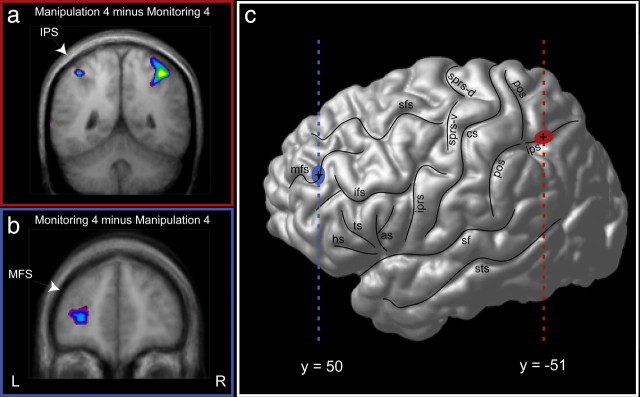
Figure 2.
Activity in the man4 minus mon4 and in the mon4 minus man4 comparisons. a, Bilateral increased activity in the IPS region in the man4 words minus mon4 words comparison. b, Increased activity in the left MDLFC in the mon4 words minus man4 words comparison. MFS, Middle frontal sulcus. c, Cortical surface rendering in standard stereotaxic space of the left hemisphere of a subject's brain. The vertical blue line indicates the anteroposterior level of the coronal section illustrated in b, and the blue circle indicates the focus of activity in the MDLFC. The vertical red line indicates the anteroposterior level of the coronal section illustrated in a, and the red circle indicates the focus of activity in the depth of the IPS. L, Left hemisphere; R, right hemisphere; as, ascending sulcus; cs, central sulcus; hs, horizontal sulcus; ips, intraparietal sulcus; ifs, inferior frontal sulcus; iprs, inferior precentral sulcus; mfs, middle frontal sulcus; pos, postcentral sulcus; sf, Sylvian fissure; sfs, superior frontal sulcus; sprs-d, dorsal branches of the superior precentral sulcus; sprs-v, ventral branches of the superior precentral sulcus; sts, superior temporal sulcus; ts, triangular sulcus.

