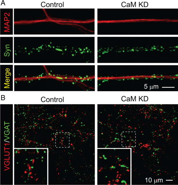
Figure 2.
CaM KD does not alter synapse density in cultured neurons. A, Representative images of neurons infected with control or CaM KD lentiviruses and immunolabeled with antibodies to MAP2 and synapsin (Syn). Synapse density was quantified by counting the number of synapsin-stained puncta per dendrite area identified by MAP2 staining using NIH ImageJ. In control condition, the average density of synapses is 5.73 ± 0.27/μm2 dendritic area; in the CaM KD condition, the average synaptic density is 5.73 ± 0.22/μm2 (n = 51 and 52 in control and CaM KD, respectively). Three batches of cultures were analyzed. B, Representative images of excitatory and inhibitory synapses in cultured neurons infected with control lentivirus or the CaM KD lentivirus. Synapses were immunolabeled with antibodies to the vesicular glutamate transporter (specific for excitatory synapses; VGLUT1) and to the vesicular GABA transporter (specific for inhibitory synapses; VGAT). Insets show magnified images from the areas indicated by dashed squares. There is no difference in the number of either VGAT or VGLUT1 puncta between control and CaM KD (supplemental Fig. 3, available at www.jneurosci.org as supplemental material). The ratios of VGLUT1/VGAT puncta are 1.01 ± 0.06 and 0.99 ± 0.06 in control (n = 41) and CaM KD (n = 43). Four different batches of cultured neurons were analyzed.

