
Figure 9.
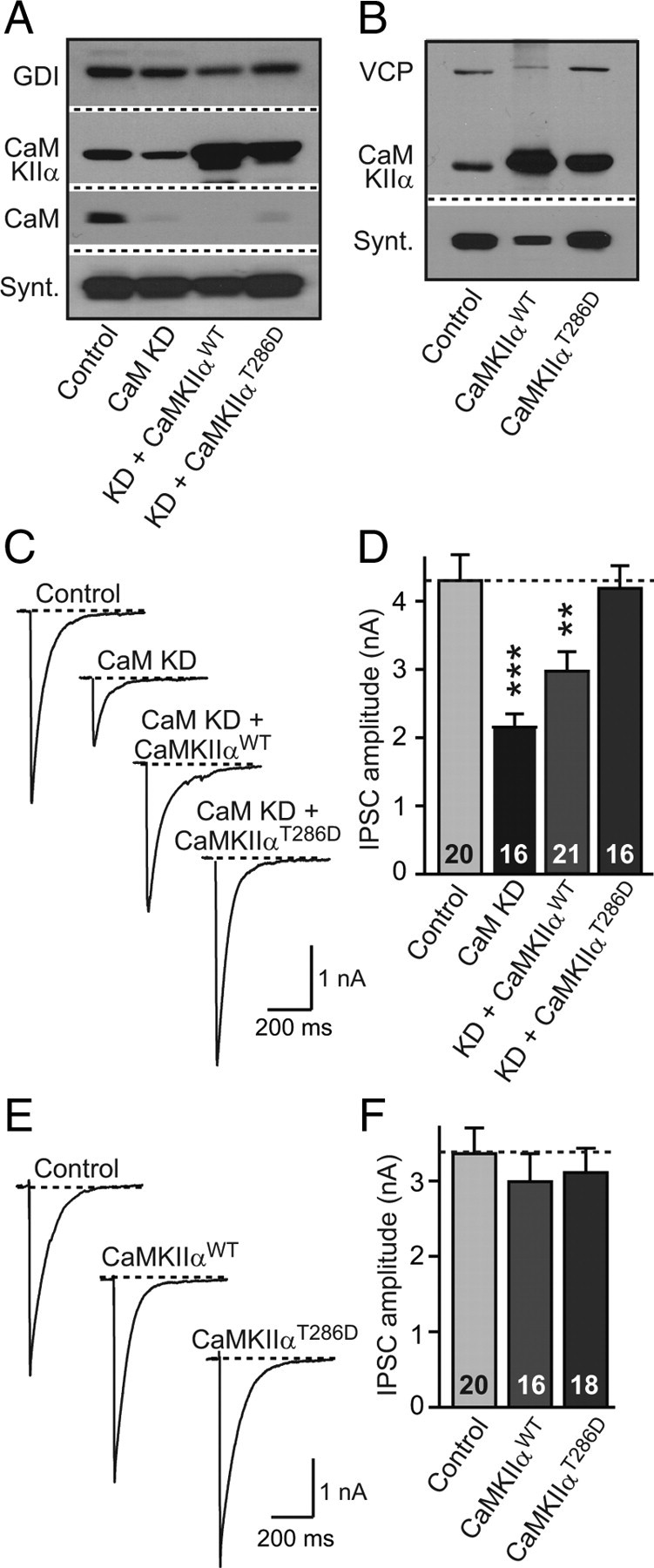
CaMKIIa overexpression rescues synaptic strength in CaM KD synapses. A, Neurons were infected with lentivirus expressing only EGFP (Control); coexpressing EGFP with CaM KD shRNAs (CaM KD); coexpressing EGFP, CaM KD shRNAs, and wild-type CaM Kinase IIα (KD + CaMKIIαWT), and coexpressing EGFP, CaM KD shRNAs, and the T286D constitutively active mutant of CaMKIIα (KD + CaMKIIαT286D). Representative immunoblots of CaMKIIα, CaM, syntaxin (Synt), and GDI (GDP-dissociation inhibitor) were used as loading control. B, Immunoblots of cultures (without CaM KD shRNAs) expressing EGFP (Control); coexpressing EGFP with CaMKIIαWT (CaMKIIαWT); and coexpressing of EGFP with CaMKIIαT286D (CaMKIIαT286D). VCP (vasolin-containing protein) was used as loading control. C, Representative traces of evoked IPSCs in CaM KD cultures with manipulations described in A. D, Summary graphs of the mean IPSC amplitudes in neurons that were manipulated as described in A. E, Representative traces of evoked IPSCs in control cultures with or without CaMKIIαWT or CaMKIIαT286D (i.e., the effect of CaMKIIα overexpression on neurons expressing normal CaM levels was investigated). F, Summary graphs of the mean IPSC amplitudes in neurons that were manipulated as described in E. Data are means ± SEM; numbers in bars indicate number of cells analyzed in at least three independent experiments; statistical significance was calculated by Student's t test. **p < 0.01, ***p < 0.001.