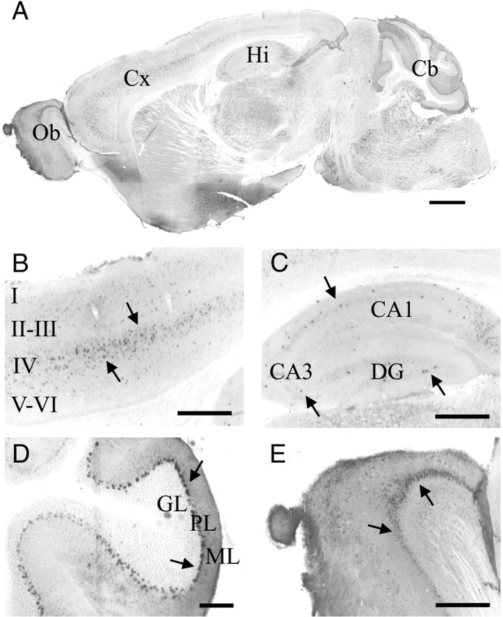
Figure 5.

Histological detection of LacZ in the brain of adult gat1(5.7m)lacz mice. A–E, Sagittal brain sections from gat1(5.7m)lacz transgenic mice were stained by anti-LacZ antibodies. Shown are images of whole brain (A), cerebral cortex (Cx; B), hippocampus (Hi; C), cerebellum (Cb; D), and olfactory bulb (Ob; E). A–E, Arrows denote immunopositive cells. Scale bars: A, 1 mm; D, 100 μm; B, C, E, 400 μm.
