
Figure 8.
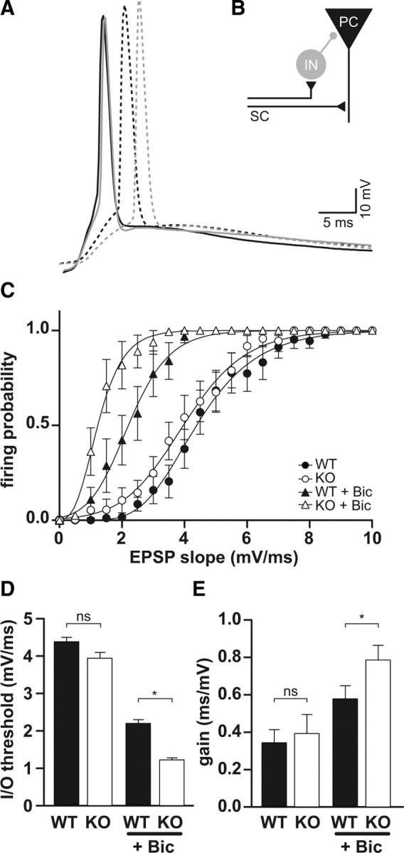
Feedforward inhibition is increased in Clcn2−/− animals. A, Sample traces of evoked EPSP in WT (black) and KO (gray) CA1 neurons in the absence (solid) and presence of bicuculline (dashed). B, Schematic diagram of disynaptic feedforward inhibition. SC, Schaffer collateral; IN, interneuron; PC, pyramidal cell. C, Neuronal I/O function. Firing probability plotted against the EPSP slope in the absence (circles) and presence (triangles) of bicuculline (Bic) for WT (filled symbols) and KO (open symbols). In the absence of bicuculline, I/O functions for WT and KO were not significantly different (n = 16 for WT; n = 16 for KO), whereas in the presence of bicuculline, the I/O function for KO was shifted more to the left relative to WT and had a steeper slope. D, E, Average gain and threshold for I/O functions shown in C. Error bars represent SEM in all graphs.