
Figure 2.
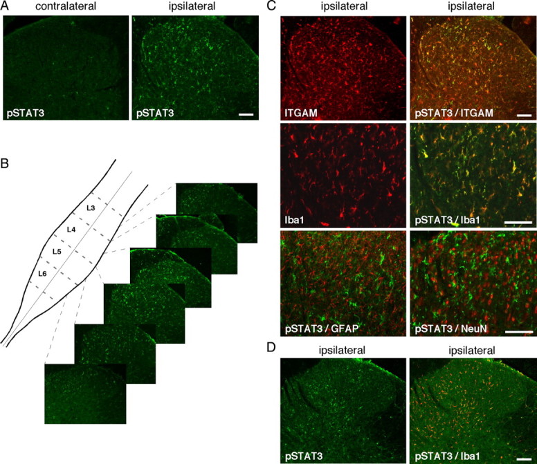
Activation of the JAK/STAT3 transduction pathway. Unilateral CCI of the rat sciatic nerve resulted in the accumulation of the active, phosphorylated form of STAT3 (pSTAT3–Tyr705, in green) 2 d later in numerous cells of the superficial and medial laminae (I–IV) of the dorsal spinal cord, on the side that is ipsilateral to the lesion. A, pSTAT-IR was almost undetectable in the contralateral side of the dorsal spinal cord of CCI rats. B, pSTAT-IR was spatially distributed from approximately the mid-L6 segment to the end of the L4 segment of the spinal cord lumbar enlargement. C, Similarly, CCI injury induced microglial activation mainly in the ipsilateral side of the dorsal spinal cord, as indicated by specific microglial markers ITGAM or Iba1 (in red). Double-labeling experiments for pSTAT3 (in green) with either ITGAM or Iba1 revealed a large colocalization of pSTAT3 with both microglial markers. Colabeling with astrocytes marker GFAP antibodies showed almost no pSTAT-IR (in green) in astrocytes and weak pSTAT3-IR signal in only a very few neurons stained with NeuN antibodies. D, One week after CCI surgery, pSTAT3 labeling (in green) was still detectable but weaker than at 2 d after injury, in the ipsilateral dorsal spinal cord and was mainly present in microglial cells labeled with Iba1-IR (in red). Scale bars, 200 μm.