
Figure 1.
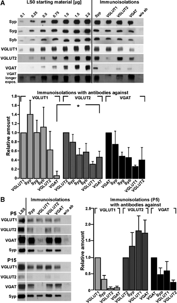
Comparison of the amounts of abundant vesicular proteins and transmitter transporters on SVs immunoisolated from adult or juvenile rat brains by antibodies against VGLUT1, VGLUT2, and VGAT. A, Immunoisolated vesicles (adult brains) were obtained as given in Materials and Methods and detection was performed with the antibodies indicated or without antibody (w/o ab). Detection of the vesicular proteins indicated on the left side was performed using the respective polyclonal rabbit antibodies. A standard curve from the starting material (LS0) was performed in parallel to calculate the respective protein amounts in the individual immunoisolates. Relative amounts of Syp, Syg, and Syb as well as of VGLUT1, VGLUT2, and VGAT were quantified from three different immunoisolation experiments using standard curves performed from the initial LS0 fraction. Note that VGLUT2-immunoisolated vesicles harbor more VGAT than VGLUT1 isolates (*Student's t test, p < 0.03). B, Transporter-specific immunoisolated vesicles were obtained as given in methods using lysed synaptosomes (LS0) from rat brains at postnatal days 5 and 15. The amount of VGAT on VGLUT2-immunoisolated vesicles was the highest at P5. Relative amounts of Syp, VGLUT1, VGLUT2, and VGAT were quantified from three different immunoisolation experiments using P5 brains. Standard curves from the initial LS0 fraction of P5 brains were performed as in B (data not shown); the LS0 fraction presented in the graph corresponds to 4 μg of protein. Blots were developed for 30 s or 3 min (longer exposure).