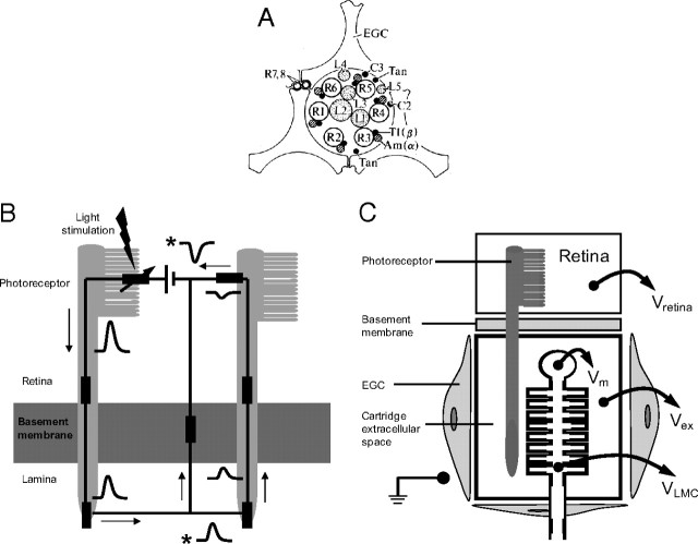
Figure 1.
Schematics showing the basic cellular and electrical structure of a lamina cartridge in the first optic neuropile of the fly. A, Transverse section through the synaptic zone of a cartridge showing the positions of the major cellular elements (redrawn after Shaw, 1981). The ring of axon terminals of the six achromatic photoreceptors coding the same image pixel (R1–R6) form ∼1300 tetradic output synapses, within which the three LMCs L1, L2, and L3 act as postsynaptic elements. The smaller elements are the two chromatic photoreceptors R7 and R8, the interneurons L4, L5, T1, and C2, and the lamina amacrine cells Am(α). Three epithelial glial cells (EGC) form a sheath around the cartridge that encloses the extracellular space and restricts diffusion between neighboring cartridges (redrawn after Shaw, 1981, 1984). B, Basic electrical circuit showing how current leaving photoreceptor terminals depolarizes the extracellular space in a lamina cartridge (redrawn after Shaw, 1975). The left-hand side of the circuit shows light-gated current entering the photoreceptor soma in the retina and leaving via its axon terminal in the lamina. To complete the circuit, the current passes from lamina extracellular space back to retina and, by crossing a glial resistance barrier at the basement membrane, depolarizes the lamina extracellular space. This depolarization can also drive return current through neighboring, less-stimulated photoreceptors (right-hand side). The waveforms indicate voltage responses to a flash of light at different locations in the circuit, all of which are measured relative to a reference electrode in the hemolymph. Responses recorded from extracellular space are denoted by an asterisk. The rest represent intracellular recordings. C, A schematic of the projection of a photoreceptor from the retina to a postsynaptic LMC in a lamina cartridge. The photoreceptor axon penetrates the resistance barrier at the basement membrane and enters the cartridge, which is a compartment bounded by EGCs. The extracellular potential within this compartment, Vex, biases the membrane potentials of cells within it. As shown for an LMC, the transmembrane potential Vm is the intracellular potential VLMC, recorded relative to a common reference minus the Vex recorded relative to the same reference. In our experiments, the reference was either the earth shown in the body cavity or, more frequently, the extracellular potential in the retina, Vretina (see text for further details).

