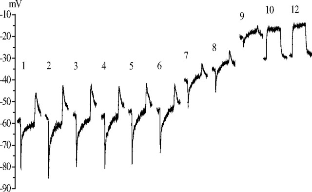
Figure 2.
The effect of DMSO injection on the membrane potential recorded from a dark-adapted LMC. After penetration by a DMSO-filled micropipette, the dark resting membrane potential of the LMC and its response to a 300 ms light step (106 photons/s) were measured at 1 min intervals. This series of responses is plotted as a series with the time after penetration in minutes above each response. Each response waveform is the average of three responses delivered 0.5 s apart and is positioned with its initial baseline at the prevailing dark resting potential. DMSO injections were started after measuring the response at 1 min, and they were performed by means of −0.25 nA current pulses lasting 300 ms, repeated with 1 s intervals. The original dark resting potential of the cell was −55 mV. During the first 5 min the on-response reduced from 40 to 30 mV without a significant change in the dark resting potential. After this, the dark resting potential depolarized by 20 mV, and a clear depolarizing plateau appeared in the response. After 10 min, the hyperpolarizing response of the LMC disappeared, leaving just the depolarizing response. The reason for the reduction of the dark resting potential in this recording between the 9th and 10th minutes was not clear, but this was not a common feature in the DMSO experiments. The final dark resting potential, indicative of the lamina extracellular space, was −32 mV.

