Figure 3.
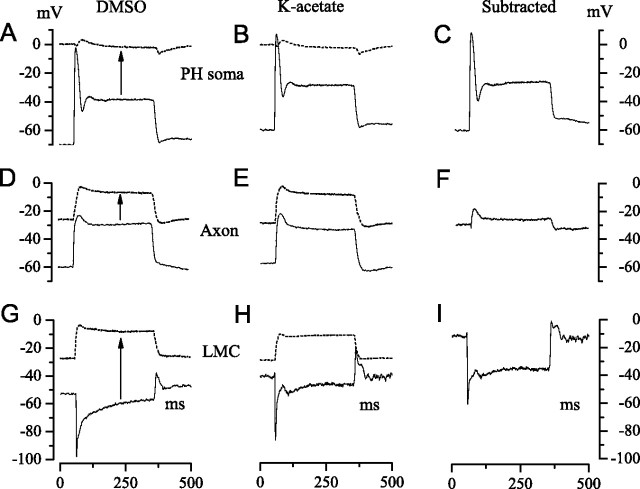
A summary of our experimental approach to recording the FP from extracellular space and determining its effect on the transmembrane potentials of photoreceptors and LMCs. A, B, D, E, G, H, The solid traces show intracellular recordings from a photoreceptor soma in the retina (A, B), and from a photoreceptor axon (D, E) and an LMC (G, H) in lamina cartridges. Membrane potentials were recorded relative to a common reference. Light responses were evoked by 300 ms pulses of 106 photons/s delivered at 1 s intervals. Two approaches were used to determine the extracellular FP (dashed traces) at the sites of these intracellular recordings. Cells were penetrated with electrodes containing DMSO (left-hand column), identified from properties recorded immediately after penetration (e.g., solid traces), and then permeabilized with DMSO to record the FP (dashed traces). In the second approach (middle column), a micropipette filled with K-acetate was used to record intracellularly from an axon or an LMC in a cartridge, and then repositioned to record from extracellular space close to the site of the intracellular recording. C, F, I, Subtraction of the extracellular record from the intracellular record gives the transmembrane potential (right column). Each trace is the average of three responses.

