Figure 5.
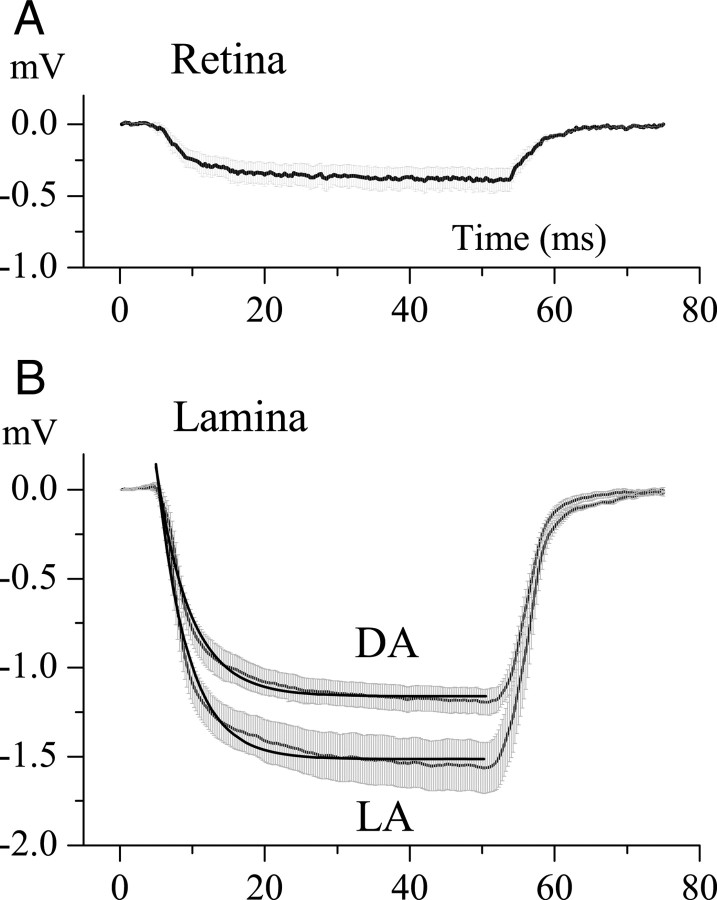
The change in potential produced by injecting current into the extracellular space of the retina and the lamina. A, Polarization of the retinal extracellular space by a −0.25 nA current pulse lasting 50 ms. The charging curve has a time constant ≈5.0 ms, and the amplitude of response at the end of the pulse indicates a resistance of 1.9 MΩ. Data were averaged over five experiments; range bars show the SE. B, Polarization of the lamina intracartridge space under dark-adapted (DA) and light-adapted (LA) conditions. Data obtained are as in A and were averaged for >10 experiments for DA and 6 experiments for LA conditions. Range bars show the SE. The dark thick lines are first-order exponentials that indicate time constants of 4.0 ms for DA conditions, and 4.2 ms for LA conditions. Assuming a simple RC circuit model for the extracellular compartment, this yields resistances of 6.0 ± 0.45 MΩ for DA conditions and 7.8 ± 0.78 MΩ for LA conditions, and apparent capacitances of 0.71 nF (DA) and 0.51 nF (LA).

