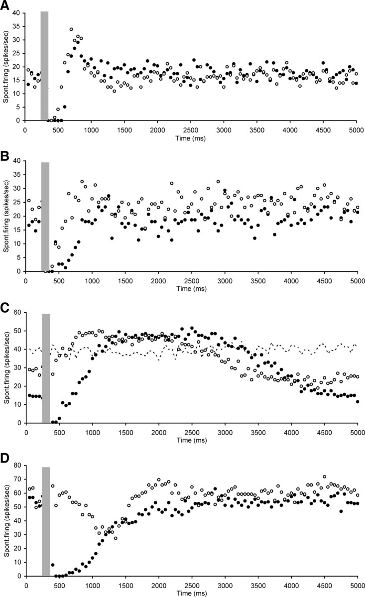
Figure 7.

Scatter plots of spontaneous firing of hyperactive CNIC neurons with electrical stimulation of the OCB (gray bar), recorded both before (dark circles) and after (open circles) strychnine perfusion in the cochlea. Illustrations of neurons in which the immediate suppression observed after OCB stimulation was not completely abolished by cochlear strychnine perfusion but its duration was reduced. In C also indicated is the spontaneous firing rate without OCB stimulation or strychnine perfusion (dotted line). A, Neuron CF 8.5 kHz, threshold 43 dB SPL. B, Neuron CF 9.2 kHz, threshold 67 dB SPL. C, Neuron nonresponsive to sound. D, Neuron CF 9 kHz, threshold 61 dB SPL. Stimulation parameters: 100 ms duration, 0.1 ms pulses, 300 Hz. Stimulation strength in A and D was 250 μA and in B and C 300 μA.
