
Figure 3.
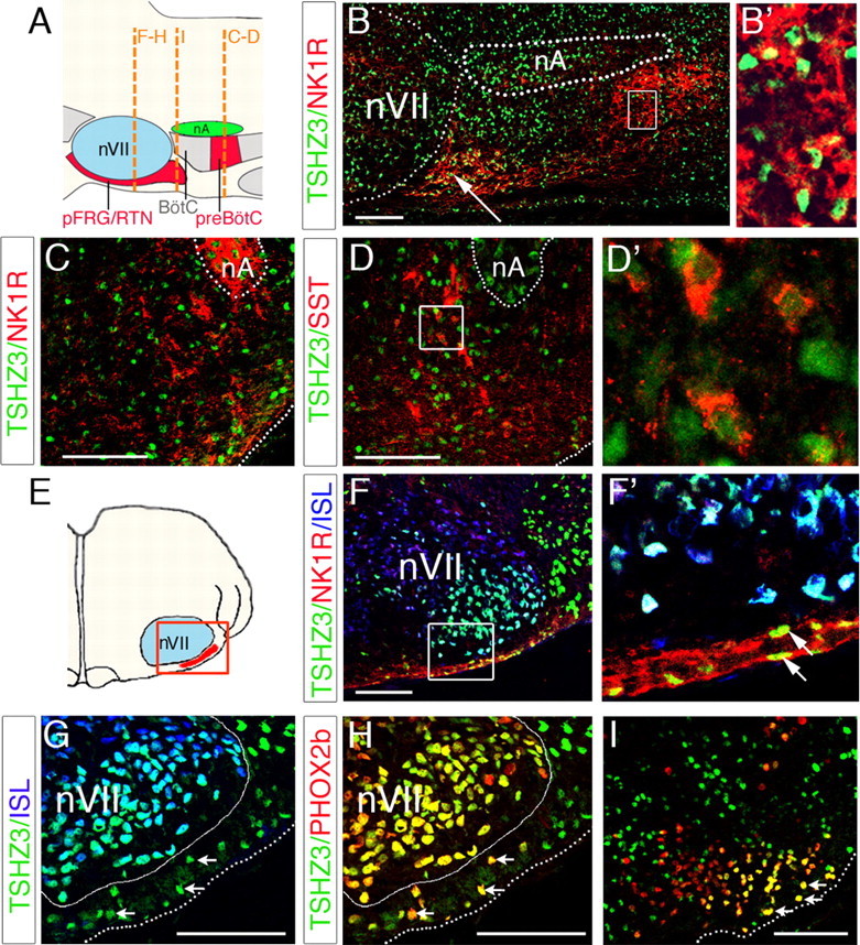
Expression of TSHZ3 in embryonic parafacial region and in preBötC. A, Schematic parasagittal view of the medullary ventral respiratory column indicating the position of the pFRG/RTN and the preBötC relative to facial nucleus (nVII) and nA, respectively. BötC, Bötzinger complex. The dashed lines show the position of the transverse sections through the nVII (F–H), immediately caudal to nVII (I) and in the preBötC (B–D). B, Parasagittal section crossing the lateral part of the nVII at E15.5. TSHZ3 is expressed in the nVII as well as in NK1R-positive cells located caudally to the nVII (white arrow), and the dotted lines indicate the position of the nVII and the nA. TSHZ3+ cells are also found in the preBötC. The inset shows that TSHZ3+ cells expressed NK1R. C, Transverse section of an E15.5 hindbrain showing expression of TSHZ3 in the NK1R-positive neurons of the preBötC. D, Transverse section of E18.5 hindbrain showing expression of TSHZ3 and SST; the dotted line delineates the nA. D′, Magnification of TSHZ3-positive cells from the box in D. E, Schematic transversal view indicating the position of sections in F–H. F, Coexpression of TSHZ3 and NK1R in cells located ventrally to the nVII, a structure immunopositive for ISL (blue). F′, Magnification of box in F. G, H, Section through the E15.5 hindbrain, used for detection of TSHZ3 and ISL (G) and TSHZ3 and PHOX2b (H). The white arrows indicate TSHZ3+ cells expressing PHOX2b in H; these same cells do not express ISL (G, white arrows). A white line delineates nVII, and a dotted line marks the ventral medullary surface. I, Transverse section performed immediately caudal to the nVII at E15.5, showing expression of TSHZ3 in the most laterally located PHOX2b+ cells (arrows). Scale bars, 100 μm.