
Figure 1.
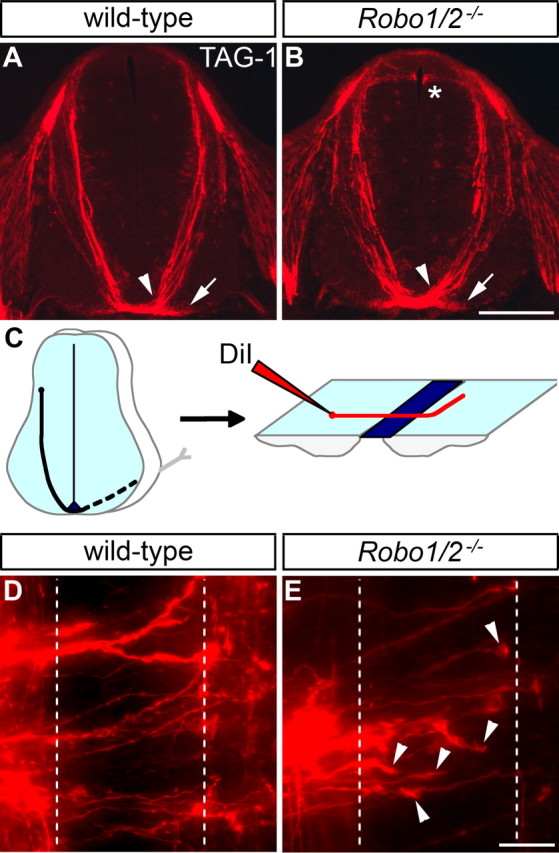
Commissural axon guidance in mice lacking Robo1 and Robo2 is affected less severely than in Slit1/2/3 mutants. A, B, Transverse sections of E11.5 spinal cords from wild-type and Robo1−/−; Robo2−/− mice were stained for TAG-1 to label commissural axons as they grow ventrally toward the floor plate and cross to the contralateral side. Commissural axons are tightly fasciculated as they approach and cross the midline (arrowhead) in both wild-type (A) and Robo1/2 mutant (B) embryos. TAG-1 staining in the medial VF (arrow) is also observed in both genotypes. TAG-1-positive sensory afferents misproject into the dorsal spinal cord of Robo1/2 mutants (*), which is not seen in wild-type. C, A schematic drawing of commissural axon projection toward and across the floor plate, as visualized by DiI injection in an open-book preparation. D, E, Open books of E12.5 wild-type and Robo1−/−; Robo2−/− embryos were injected with DiI as shown in C. In wild-type mice (D), axons project across the ipsilateral (left dashed line) and contralateral (right dashed line) floor plate borders and then turn anteriorly (up). In mice lacking Robo1 and Robo2 (E), many commissural axon growth cones are observed in the floor plate (arrowheads). Injection sites with five or more growth cones in the floor plate are more frequent in Robo1/2 mutants (45% of 42 injections in 5 embryos) than in wild type (18% of 40 injections in 5 embryos). Scale bars: A, B, 200 μm; D, E, 25 μm.