
Figure 2.
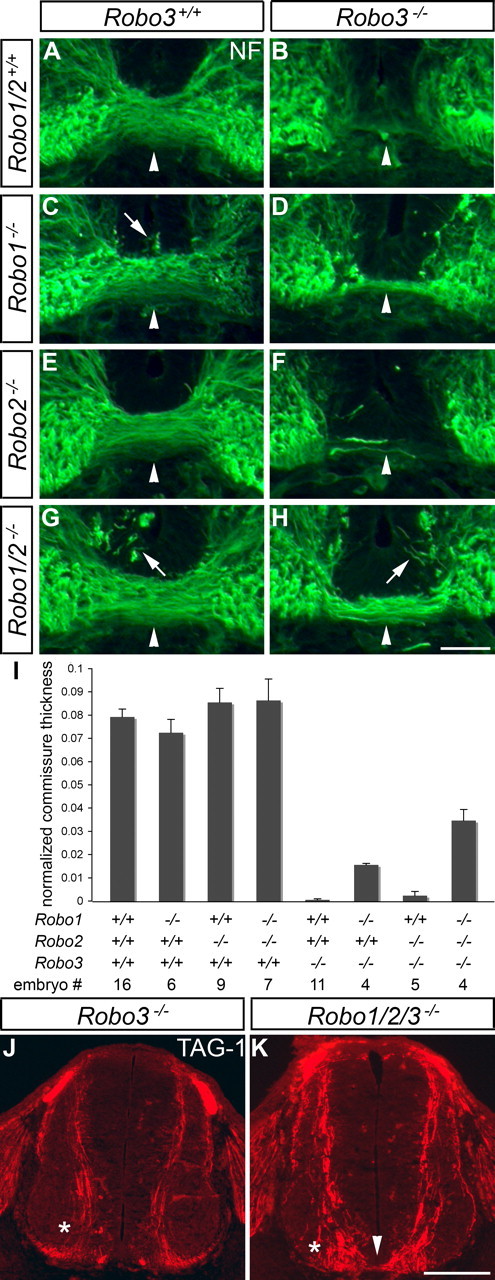
Loss of Robo1 and Robo2 does not fully rescue midline crossing in Robo3 mutants. A–I, Transverse sections of E11.5 spinal cords from mice with the indicated genotypes were stained for neurofilament (NF), and the thickness of the main commissural bundle was quantified (see Materials and Methods). In wild-type embryos (A), axons form a compact fascicle (arrowhead) under the floor plate. In Robo3 mutant mice (B), axons fail to cross the midline. In mice lacking Robo1 (C), some axons invade the floor plate (arrow), but the main commissural fascicle is comparable in size with wild-type embryos. Loss of Robo1 in a Robo3 mutant background (D) partially rescues midline crossing (p = 0.009 when compared with Robo3−/−; p < 0.0001 when compared with wild type). The ventral commissure in Robo2−/− mice (E) looks similar to wild-type mice, and loss of Robo2 in a Robo3−/− background (F) restores midline crossing in a few cases (not statistically significant) but less than observed in Robo1−/−; Robo3−/− embryos. Axons are invading the floor plate (arrow) in mice lacking Robo1 and Robo2 (G), but the commissure is similar in size to wild-type embryos. In Robo1−/−; Robo2−/−; Robo3−/− mice (H), the commissure is thicker than in mice lacking Robo1 and Robo3 (p = 0.0069) but is still significantly thinner than in wild-type embryos (p < 0.0001). Error bars in I indicate SEM. J, K, TAG-1 staining of E11.5 spinal cord sections labels commissural axons as they approach and cross the midline. In Robo3 mutants (J), all commissural axons stay ipsilaterally (*). In Robo1−/−; Robo2−/−; Robo3−/− embryos (K), some axons cross the floor plate (arrowhead), whereas others veer away from the midline and stay on the ipsilateral side (*). Scale bars: A–H, 50 μm; J, K, 200 μm.