
Figure 3.
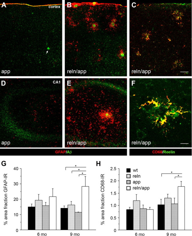
Increased inflammatory responses in reln/app double transgenic mice. Low-magnification images of double immunofluorescence staining of cortical (A–C) and hippocampal (D–F) brain sections obtained from 9-month-old single mutant app and double mutant reln/app mice. A, B, D, E, Pronounced increase in GFAP-positive astrocytes (red) selectively associated with amyloid-β plaques (anti-Aβ1-40/42 antibody; green) was found in reln/app compared with app subjects. C, F, A similar increase in reactive microglia was evident in the neocortex (C) and hippocampus (F) of 9 month reln/app mice. Note the selective accumulation of CD68-positive microglia (red) with Reelin-immunoreactive plaques (green). G, Quantification of the area fraction of GFAP-immunoreactive astrocytes revealed a main effect of genotype (F (3,20) = 4.4; p = 0.016) and significant differences between reln/app versus wt (p = 0.022), reln/app versus reln (p = 0.043), and reln/app versus app (p = 0.010) at 9 months of age. H, Similar differences emerged between reln/app versus wt (p = 0.039) and reln/app versus app (p = 0.046) subjects for the CD68 area fraction. Values are given as mean ± SEM. *p < 0.05 (comparison with reln/app subjects), statistical significance based on Fisher's LSD post hoc analysis. Scale bars: C, 50 μm; F, 10 μm.