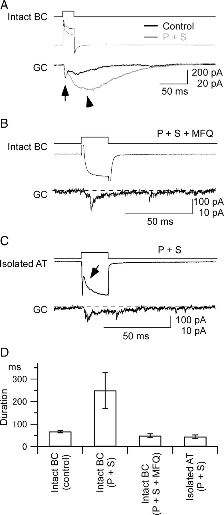
Figure 6.

Synaptic transmission from Mb1-BC to GC. A, Long-lasting EPSC evoked by a brief depolarization of Mb1-BC. Paired recordings from an intact Mb1-BC (intact BC) and a postsynaptic ganglion cell (GC) in the dark-adapted retina. A depolarizing pulse (from −60 to −10 mV for 20 ms) (top) applied to the intact BC evoked an outward current (middle) in the intact BC and an EPSC (bottom) in the GC voltage clamped at −60 mV. Recordings were performed in the control solution (Control, black traces), and then in the presence of picrotoxin (200 μm) and strychnine (10 μm) (P + S, gray traces). The pipette for the intact BC was filled with the Cs+-based pipette solution and that for GC with the Cs+-based pipette solution containing QX-314 (5 mm). The evoked EPSC consisted of the fast (arrow) and slow (arrowhead) components. B, Effects of a gap junction blocker on the synaptic transmission from Mb1-BC to GC. Recordings were made from a pair of an intact BC and a postsynaptic GC in the dark-adapted retinal slice preparation, which was preincubated for >30 min with MFQ (10 μm), picrotoxin (200 μm), strychnine (10 μm), and L-AP4 (100 μm). The pipette for the intact BC was filled with the Cs+-based pipette solution and that for the GC with the Cs+-based pipette solution containing QX-314 (5 mm). A brief depolarization (top; from −60 to −10 mV for 50 ms) of the intact BC evoked a Ca2+ current (middle) in the intact BC and a transient EPSC (bottom) in the GC. The dotted line indicates the basal current at the holding potential of −60 mV. C, Recordings from a pair of an isolated axon terminal (isolated AT) of Mb1-BC and a postsynaptic GC in the dark-adapted retina. Both were voltage clamped at −60 mV. A brief depolarization (top; to −10 mV for 20 ms) of the isolated AT evoked a Ca2+ current (middle) in the isolated AT and an EPSC (bottom) in the GC. The arrow indicates the proton feedback. The dotted line indicates the basal current at the holding potential of −60 mV. The external solution contained picrotoxin (200 μm), strychnine (10 μm), and TTX (0.5 μm), and the Cs+-based pipette solution. D, Pooled data of duration of the EPSCs evoked in the postsynaptic GCs. Duration was defined as the response width of 10% of the peak amplitude. Data were obtained from five pairs of an intact BC and a GC in control condition [Intact BC (control)], five pairs of an intact BC and a GC in the solution containing picrotoxin and strychnine [Intact BC (P + S)], five pairs of an intact BC and a GC in the solution containing picrotoxin, strychnine, and mefloquine [Intact BC (P + S + MFQ)], and three pairs of an isolated AT and a GC in the solution containing picrotoxin and strychnine [Isolated AT (P + S)].
