
Figure 2.
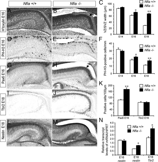
Delayed development of ventricular zone progenitors in Nfia mutants. A, B, Coronal paraffin sections of E16 wild-type and Nfia−/− brains stained with hematoxylin. The ventricular zone was wider in the mutant (arrow in B). C, Quantification of ventricular zone width at E14, E16, and E18 revealed that the ventricular zone was significantly wider in the Nfia mutants compared to controls at both E16 and E18. D, E, PH-H3 immunohistochemistry on coronal paraffin sections of E16 brains. F, Quantification of the number of PH-H3-positive cells revealed significantly more mitotic cells in the mutant ventricular zone at both E16 and E18. G, H, Immunohistochemistry against Pax6 on coronal brain sections at E16. K, Cell counts demonstrated that there were significantly more Pax6-positive cells in the ventricular zone of Nfia mutants (arrow in H) compared to controls. No difference in the number of Tbr2-positive cells within the hippocampus was detected at E16. I, J, Tbr2 immunohistochemistry at E18 revealed higher expression of Tbr2 expression in Nfia−/− hippocampi in late gestation. L, M, Expression of nestin in wild-type (L) and Nfia−/− (M) brains at E18. N, qPCR on wild-type and Nfia−/− hippocampal tissue revealed a significant increase in both nestin and Tbr2 mRNA levels in the mutant at E18. *p < 0.05, **p < 0.001, Student's t test. Scale bar (in M): A–H, 250 μm; I–M, 300 μm.