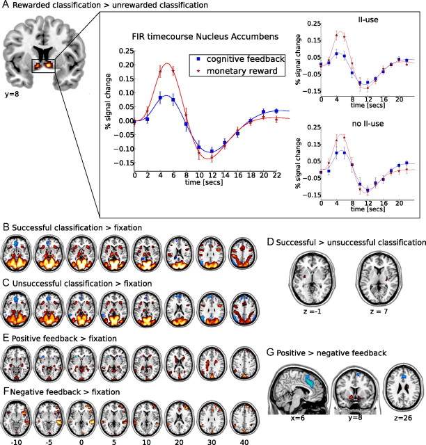
Figure 4.
fMRI results. A, Activation in the contrast of monetary reward minus cognitive feedback during stimulus presentation. The time course represents the finite impulse response (FIR) to both monetary reward and cognitive feedback during stimulus presentation, extracted using MarsBar and an anatomical ROI of the nucleus accumbens from the Harvard–Oxford subcortical structural atlas. For each subject, individual functional ROIs within this anatomical ROI were defined based on the areas in which the main effect of stimulus presentation exceeded an uncorrected threshold of p ≤ 0.1. Error bars represent the SEM. Results of this analysis are also plotted separately for subjects with use of the optimal decision bound in at least one condition and those with no information-integration use (no II use). A significant peak activation difference between the task conditions is only observed in the group of subjects with information-integration use (II use). B, C, E, F, Activations (yellow to red) and deactivations (white to blue) for contrasts against fixation. B, Successful categorization minus fixation. Activations are observed in occipital and parietal cortices, as well as in subcortical areas. C, Unsuccessful categorization minus fixation. Activations are mainly observed in occipital and parietal cortices. D, Activations in the contrast of successful minus unsuccessful categorization. No voxel showed higher activations for unsuccessful categorization, whereas bilateral clusters of higher activation during successful categorization were observed in the putamen. E, Positive feedback minus fixation. Both the caudate nuclei and the hippocampi are activated. F, Negative feedback minus fixation. Activations include the rostral cingulate zone and right prefrontal areas. G, Activations in the contrast of positive minus negative feedback. Voxels that were more activated during the processing of negative feedback include the RCZ and anterior insula, whereas voxels more activated during the processing of positive feedback are observed in the nucleus accumbens and right caudate body. All maps are thresholded at a level of pFWE < 0.05. Left hemisphere is presented at the left.

