
Figure 5.
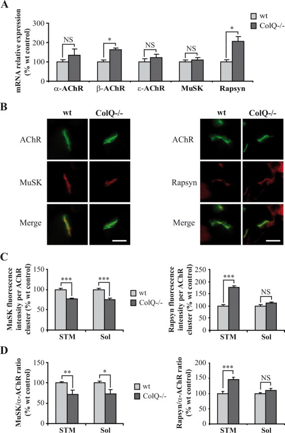
ColQ regulates AChR and rapsyn mRNA levels and MuSK membrane insertion at neonatal NMJs. A, Quantification of AChR subunits, MuSK, and rapsyn mRNAs on wt and ColQ−/− P7 gastrocnemius muscles by real-time RT-PCR. Levels of mRNAs are represented as relative expression (2−ΔCt versus reference gene × 100). β-AChR subunit and rapsyn mRNA levels relative to GAPDH mRNAs were higher in ColQ−/− mice compared to wt mice whereas other AChR subunits and MuSK mRNAs were not significantly increased. B, AChR, MuSK and rapsyn clusters labeled respectively with Alexa 488 α-BTX, MuSK and rapsyn antibodies on cross-sections of STM muscle from wt and ColQ−/− P7 mice. C, D, Quantification of MuSK and rapsyn fluorescence intensities from projection of confocal stacks. MuSK fluorescence intensity per AChR cluster was decreased in the absence of ColQ whereas rapsyn was increased (C). STM and Sol synapses from ColQ−/− mice displayed lower MuSK-to-α-AChR fluorescence ratio but higher rapsyn-to-α-AChR ratio compared to wt synapses (D). Data are presented as percentage of wt control ± SEM from five mice for mRNA analysis (A) and from three mice for immunofluorescence analysis (n ≥32 NMJs for C, D). *p < 0.05; **p < 0.01; ***p < 0.001; NS, not significant; unpaired Mann–Whitney's U test for mRNA analyses and unpaired Student's t test for immunofluorescence analyses. Scale bars, 20 μm.