
Figure 1.
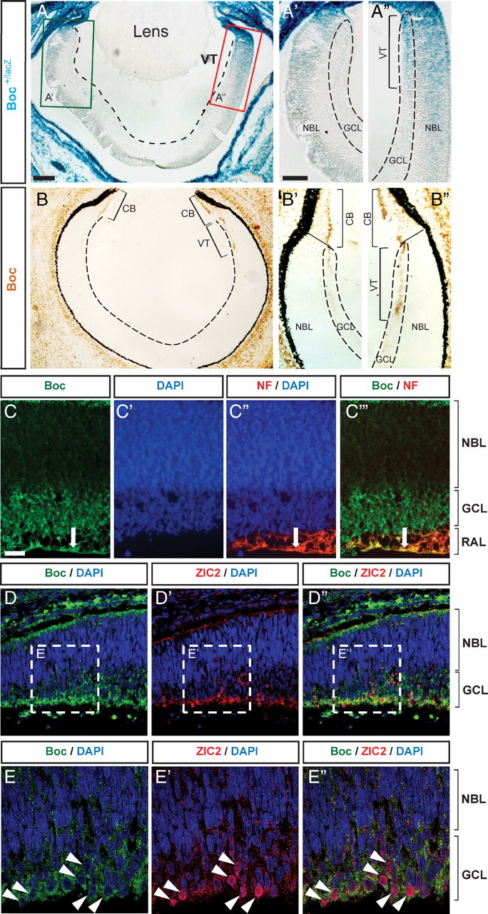
Boc is preferentially expressed in ipsilateral RGCs. A–B″, Boc expression detected by β-gal histochemistry in Boc +/lacZ mice (A–A″) and immunohistochemistry with an antibody against Boc (goat anti-Boc) in E15 horizontal section of the mouse retina (B–B″). Both methods show a strong enrichment of Boc in the VT retina as shown in A″ and B″. C–C‴, Boc (green, rabbit anti-Boc antibody) is expressed by RGC cell bodies in the GCL and is present in the RAL of the VT. Boc colocalizes with neurofilament (NF) on the axons of RGCs in the RAL (arrow; C–C‴) (horizontal sections of E17 rat retina). D–D″, Zic2-positive cells (red) colocalize with Boc (green, goat anti-Boc antibody) in the VT crescent of E15 mouse retina (horizontal sections). Higher magnification of the boxed regions from D and D″ shows the nuclear localization of Zic2 surrounded by membranous expression of Boc in the GCL (E–E″). CB, Ciliary body. Scale bars: A, 100 μm; A′, A″, 50 μm; C–C‴, 20 μm.