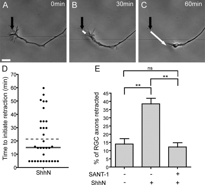
Figure 2.
Shh induces the rapid retraction of a subset of RGC axons. E17 rat retinal explants were cultured for 24 h, which allows RGC axons to elongate long distances out of the explant. A–C, Time-lapse images showing the retraction of an RGC axon following addition of 10 nm ShhN. A, Morphology of an RGC axon and growth cone at the time of ShhN addition. The black arrow points to the initial position of the growth cone. B, The growth cone starts to collapse. C, The growth cone has completely collapsed and the axon is strongly retracting. D, Time for a growth cone to initiate retraction in response to ShhN. The plot shows a sample of 33 RGC axons from four different experiments. The bar indicates the median time to initiate retraction (15 min). The dashed line indicates the mean time to initiate retraction (22 min). E, Quantification of the percentage of axons that exhibit net retraction 90 min after ShhN treatment. ShhN induces an increase in the percentage of axons with net retraction from 14.0 to 38.5% (p < 0.01). This effect is completely blocked by adding an inhibitor of Smoothened (SANT-1, 137 nm) 1 h before ShhN treatment (12.2% of RGC axons show net retraction; p < 0.01) (one-way ANOVA with Bonferronni's post-test, **p < 0.01). The mean percentage of retracted axons was quantified from at least three independent experiments for each condition, with at least four retinal explants per condition per experiment. Error bars indicate SEM. Scale bar, 10 μm.

