
Figure 1.
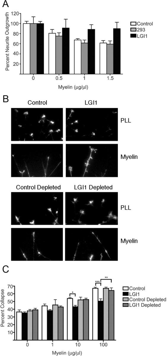
Myelin-induced growth cone collapse and neurite outgrowth inhibition is antagonized by LGI1. A, P8 rat CGNs plated on the indicated concentrations of myelin were maintained in normal media or in media conditioned by 293T cells that had, or had not, been transfected with an LGI1 expression plasmid, as indicated. B, Representative images of E13 chick DRG neurons incubated for 1 h in the presence of the indicated conditioned media then exposed to either PBS or soluble myelin (100 μg/ml) for 30 min. For depletion, media were preincubated with streptavidin agarose beads to remove metabolically biotinylated LGI1 (see Materials and Methods). C, Quantification of DRG neuron growth cone collapse. Neurons were exposed to increasing amounts of myelin in the presence of the indicated conditioned media. For A and C, statistical differences between groups was established using two-way ANOVA followed by Bonferonni post hoc tests (*p < 0.05, **p < 0.01, ***p < 0.001).