
Figure 8.
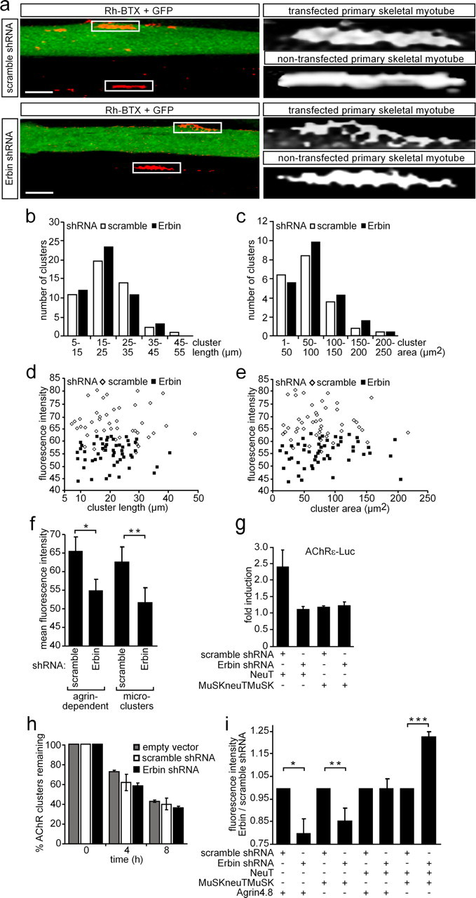
Knockdown of Erbin reduces the density of AChR aggregates. a, Primary skeletal myoblasts were transfected with shRNA (Erbin-specific or scrambled) cloned in pSuperGFPneo, differentiated to myotubes, and incubated with nerve-derived agrin. On the left, typical confocal images (compressed Z-stack) are shown. On the right, high-resolution grayscale images of AChR aggregates localized on transfected or nontransfected myotubes are shown. Note that the AChR aggregates formed in GFP-positive myotubes are less dense. Scale bar, 25 μm. b, c, Graphs represent comparisons of surface areas and lengths of AChR clusters as counted on primary skeletal myotubes after transient transfection with either scrambled or Erbin-specific shRNA (n > 65). d, e, Fluorescence intensities of AChR aggregates on primary skeletal myotubes are plotted against cluster length or surface area. Note that most of the AChR aggregates are of significantly lower density if muscle cells were transfected with Erbin-specific shRNA compared to scrambled shRNA. f, Graph summarizes data presented by d and e showing the mean fluorescence density of all analyzed AChR aggregates. Further, primary muscle cells were transfected as in a but not incubated with nerve-derived agrin for the analysis of the mean fluorescence intensities of AChR microclusters (n > 32). Note that even AChR microclusters are significantly less dense if Erbin is knocked down. Student's t test, *p < 2 × 10⁻¹¹, **p value <2.4 × 10⁻⁴. g, Primary skeletal myoblasts were transiently transfected, differentiated to myotubes, and harvested for preparation of cell extracts. Luciferase activities were measured a minimum of three independent times with an AChRε-luciferase reporter and different expression plasmids. NeuT-mediated enhancement of AChRε transcription was inhibited if concomitantly Erbin was knocked down. h, To find out whether AChR cluster stability decreased in the absence of Erbin, nerve-derived agrin was depleted from cell media at the indicated times, and remaining AChR aggregates were counted at 0 (pSuper, scramble, Erbin-specific, n = 76/67/70) or after 4 (n = 80/67/82) and 8 (n = 71/72/79) hours of agrin withdrawal and presented as percentage of remaining clusters. i, Graph summarizes the ratio of mean fluorescence densities of AChR aggregates on C2C12 cells, which were transfected as indicated. After transfection of the cells with MuSKneuTMuSK, incubation of the cells with nerve-derived agrin became no longer necessary because MuSKneuTMuSK induces formation of AChR aggregates even in the absence of agrin. Note that AChR aggregates formed after knockdown of Erbin in the presence of MuSKneuTMuSK and neuT together are even more dense than aggregates formed in the presence of Erbin. Student's t test, *p < 1.2 × 10⁻⁷, **p < 8.1 × 10⁻⁹, ***p < 1.2 × 10⁻⁸.