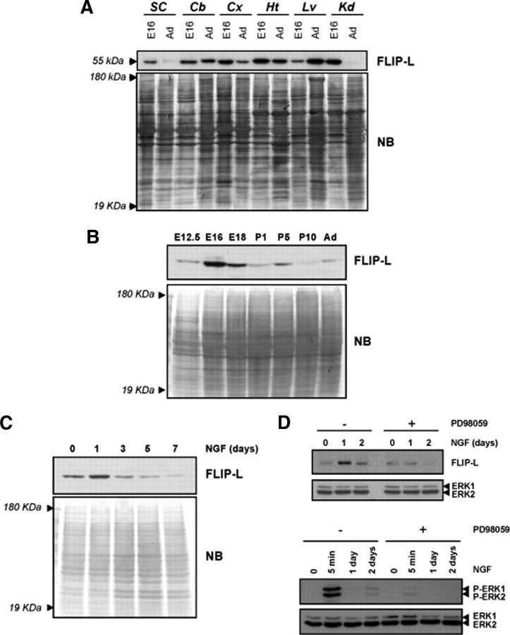
Figure 1.
FLIP-L is expressed in the nervous system and is upregulated during neuronal differentiation in vivo and in vitro. A, Embryonic (E16) and adult (Ad) mouse tissue lysates (20 μg) were analyzed by SDS-PAGE/immunoblot using anti-FLIP Dave-2 antibody (Alexis Biochemicals) (top). SC, Spinal cord; Cb, cerebellum; Cx, Cortex; Ht, heart; Lv, liver; Kd, kidney. B, Cortical brain lysates of mice at different embryonic and postnatal stages were processed, and FLIP-L immunoblot was performed. C, Time course of FLIP-L protein expression in PC12 cells subjected to NGF-induced differentiation. For loading control, the membranes were stained with Naphtol Blue (NB). D, After serum deprivation during 12 h, PC12 cells pretreated or not with 50 μm PD98059 were treated with NGF for 5 min (bottom) or 1 and 2 d (top and bottom). Total protein extracts were analyzed by Western blotting using an anti-phospho-ERK1/2 antibody (bottom) or an anti-FLIP (Dave-2) antibody for detection of FLIP-L upregulation after NGF treatment (top). Note that FLIP-L was not upregulated after 1 d of NGF treatment when cells were cultured in the presence of PD98059 (top). Membranes were stripped and reprobed with an anti-pan-ERK antibody to control protein loading.

