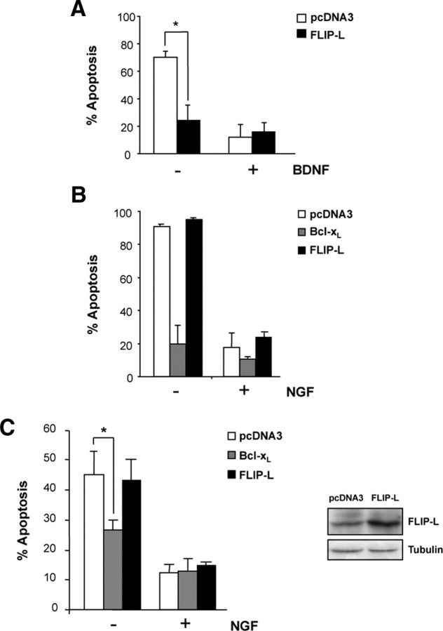
Figure 3.
A, Cultured mouse MTNs were transfected using Lipofectamine2000 with an EGFP expression plasmid together with pcDNA3 or pcDNA3–FLIP-L (FLIP-L) vectors. Cells were treated with 10 ng/ml BNDF (+) or basal medium (−) for 24 h. Percentage of apoptotic EGFP-labeled MTNs showing nuclear condensation type II was quantified using Hoechst staining. B, Ballistic transfections of cultured mouse SCG neurons with an EGFP expression plasmid together with the pcDNA3 control vector (pcDNA3), pcDNA3–FLIP-L (FLIP-L), or pcDNA3–Bcl-xL (Bcl-xL) plasmids were performed. After 24 h of incubation with 10 ng/ml NGF (+) or basal medium (−), percentage of apoptotic EGFP-labeled SCGs was quantified. C, PC12 cells stably transfected with pcDNA3, Bcl-xL or FLIP-L were treated with basal medium without serum supplemented with 100 ng/ml NGF for 3 d before trophic factor deprivation. Percentage of apoptotic cells showing type II nuclear condensation after 48 h of NGF deprivation was quantified. Significant differences are indicated (*p < 0.001, t test). Right, Total lysates were obtained and analyzed by Western blot to assess FLIP-L levels in PC12 stably transfected using anti-FLIP (Dave-2). The membrane was reblotted with anti-tubulin as a loading control.

