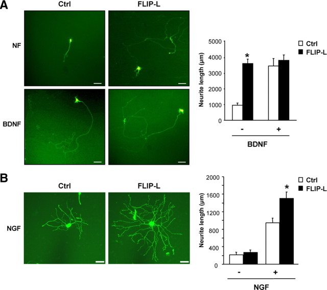
Figure 4.
A, Cultured mouse MTNs were transfected with an EGFP expression plasmid together with the control vector pcDNA3 (Ctrl) or pcDNA3–FLIP-L (FLIP-L) vectors. After 48 h of incubation with Neurobasal medium alone (NF) or supplemented with 10 ng/ml BDNF (BDNF), images were acquired and analyzed. Representative micrographs (scale bars, 100 μm) and quantification of neurite outgrowth are shown. Significant differences are indicated (*p < 0.001, t test). B, Ballistic transfections of cultured SCG neurons with an EGFP expression plasmid together with the control vector pcDNA3 (Ctrl) or pcDNA3–FLIP-L (FLIP-L) plasmids were performed 3 h after plating. After 24 h of incubation or not with 10 ng/ml NGF (NGF), EGFP-labeled SCG were visualized and digitally acquired. Representative micrographs depict the increased neurite arbor size and complexity (FLIP-L vs Ctrl), and quantification of neurite outgrowth is presented. Scale bars, 50 μm. Significant differences are indicated (*p < 0.001, ANOVA test).

