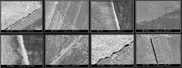
Figure 2.
(e1 and e2) A representative scanning electron microscopic image of bonded interface in control group. Notice the larger gap between dentin and restoration (×500). (f1 and f2) A representative scanning electron microscopic image of bonded interface in chlorhexidine group where gap-free interface can be observed (×500). (g1 and g2) A representative scanning electron microscopic image of bonded interface inAloe veragroup where gap-free interface can be observed (×500). (h1 and h2) A representative scanning electron microscopic image of neem group where interfacial gap can be observed (×1000)

