
Figure 3.
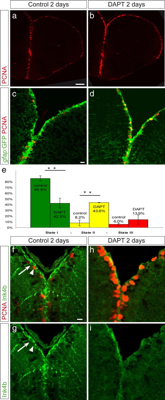
Inhibition of Notch activity triggers a state I to II switch. Vehicle-treated fish (a, c, f, g) and fish treated with 40 μm DAPT (b, d, h, i) were compared. a, b, Overview on a telencephalic cross section, as a single confocal plane, of a gfap:GFP fish stained for PCNA (red), revealing the increased density of PCNA-positive cells along the ventricular zone in DAPT-treated fish. c, d, Dorsal telencephalic ventricular zone of gfap:GFP, control (c), or DAPT-treated (d) fish, revealing an increase in state II cells (PCNA, red; GFP, green) within the GFAP-positive cell population. e, The relative proportions of states I, II, and III cells were calculated in n = 4 control brains (2845 cells) and n = 3 DAPT-treated brains (3199 cells). The difference between control and DAPT is highly significant for state I and state II cells (t test, **p < 0.001). The density of GFAP-positive cells per ventricular surface was unchanged. Error bars show the SEM (n = 4 and 3). f–i, DAPT-treated (h, i) and vehicle-treated (f, g) brains were immunostained for Ink4b (green). Ink4b is found in noncycling cells (arrow) but not in PCNA-positive cells. Ink4b expression disappeared during DAPT treatment. Scale bars: a, 50 μm; c, f, 10 μm.