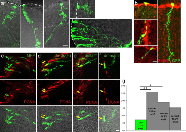
Figure 4.
In vivo transfection via lipofection and electroporation of dominant-negative constructs of the Notch pathway induces an increase of PCNA-positive cells. The ventricular zone of the telencephalon in adult fish was lipofected or electroporated with constructs in the pCS2 vector expressing eGFP (a–c) or was coelectroporated with pCS2–eGFP together with pCS2–dn-Su(H) (d), pCS2–deltastu (e), or pCS2–dn-maml1 (f). a and b depict the morphology and S100β expression (b) of transfected radial glia, which can be evaluated as individual cells. Transfected cells in c–f were assessed for their expression of PCNA (red) 2 d after electroporation; green arrows point to single GFP-positive cells, and yellow arrows point to GFP and PCNA double-positive cells. The percentage of GFP-positive cells also expressing PCNA is plotted in g for the different constructs. Note that all three dominant-negative proteins with Notch blockade activity increase cell cycle entry. In the case of dn-Su(H) and DeltaStu, this increase reaches significance (p = **0.0004 and *0.02, respectively, Fisher's exact test). n is the total number of cells counted of 5–10 brains per construct. Scale bars, 10 μm.

