
Figure 5.
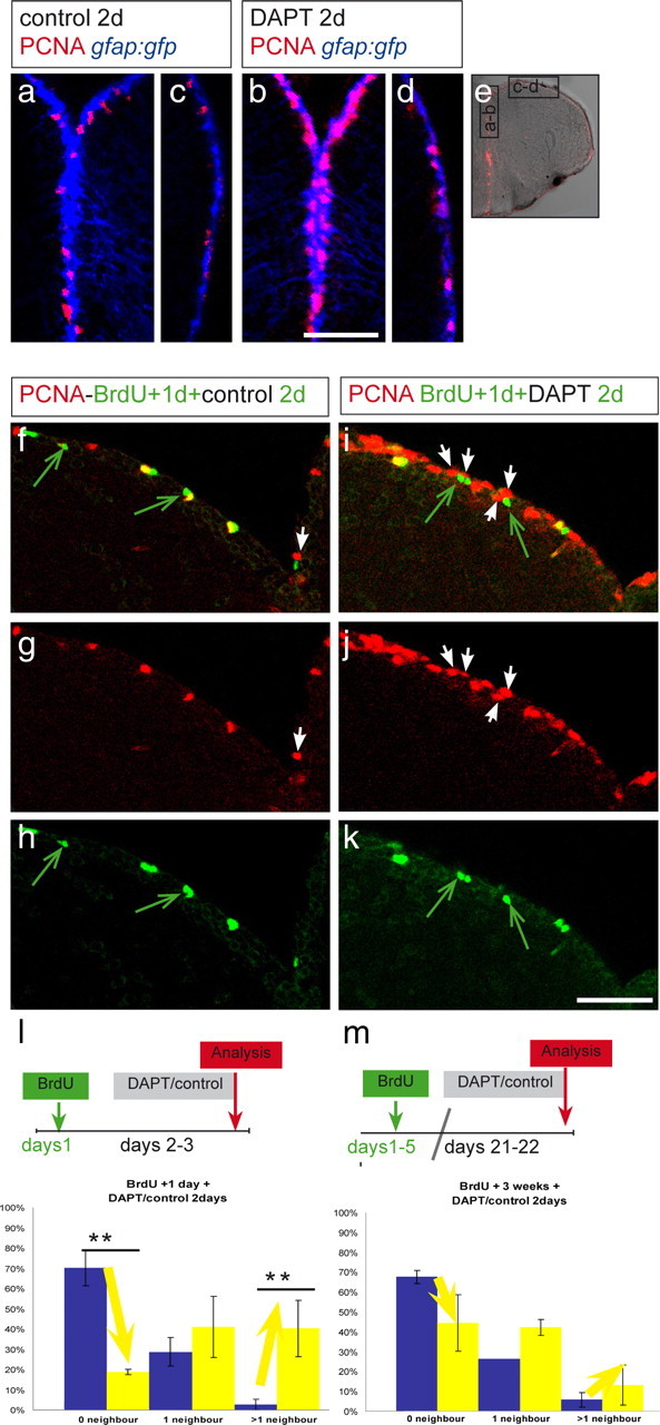
Notch maintains quiescence in cells neighboring active progenitors. DAPT-treated (b, d, i–k) or vehicle-treated (a, c, f–h) fish were compared. e, Overview of a telencephalic cross section. Black rectangles depict regions enlarged in a–d. a, b, Medial ventricular area, containing dividing progenitors (PCNA, red) under normal conditions (a) and a higher density of PCNA-positive cells after DAPT (b). c, d, Dorsolateral area of the telencephalon (as boxed in e; medial is to the top and lateral to the bottom) containing few PCNA-positive cells under normal conditions (c) and a slight increase of PCNA-positive cells after DAPT (d). The proportion of PCNA-positive cells reaches a higher level in medial regions under Notch-blocking conditions. f–k, Dorsomedial region harboring a density of PCNA-positive cells comparable with the region in a. BrdU was administered twice within 3 h, and 1 d later, the fish were treated with vehicle (f–h) or DAPT water (i–k) for 2 d. Cells neighboring BrdU-positive cells were analyzed for PCNA expression. Few BrdU-labeled cells in control brains are in contact with newly dividing, PCNA-positive cells (white arrow, f, g), and many BrdU-positive cells (some of them are still PCNA positive; yellow) have no PCNA-positive (red) neighbor (green arrows, f, h). Most BrdU-labeled cells after DAPT are surrounded by PCNA-positive cells (arrows in i–k), suggesting that Notch prevents neighbors of an already dividing cell to enter cell cycle. The amount of BrdU-positive cells is unchanged, suggesting that the cell cycle speed has not been changed by the DAPT treatment. l, m, BrdU-labeled cells, counted in two brains for each condition, were categorized in three groups according to their neighbors: no dividing neighbor, one dividing neighbor, and more than one dividing neighbor. The proportion of cells belonging to each category is represented, and the error bars represent the SEM, in which n is number of brains. The blue bar of each category represents vehicle-treated and the yellow bar DAPT-treated animals. l, In DAPT-treated animals, the proportion of BrdU-positive, 3-d-labeled cells without dividing neighbor is decreased, whereas the proportion of BrdU-labeled cells with more than one neighbor is increased (one-way ANOVA for repeated measurements; 6 sections from 2 control brains and 4 sections from 2 DAPT brains; **p < 0.01 for 0 neighbors, **p = 0.02 for 2 or more neighbors). m, Fish were injected for 5 d with BrdU and treated 3 weeks after the last injection with DAPT or with DMSO. The neighbors from these BrdU-positive, 3-week-labeled cells did not reveal a significant difference after DAPT treatment compared with control treatment. The nonsignificant changes observed are probably attributable to the general increase of dividing cells along the VZ. Scale bars: in a for a–d, in f for f–k, 100 μm.