
Figure 7.
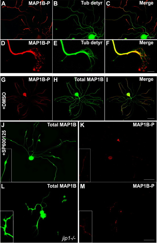
JNK inhibition affects MAP1B phosphorylation in regenerating neurons. A–C, In control DRG neurons, distal parts of neurites are enriched in MAP1B-P and characterized by low levels of detyrosinated tubulin (representing stable microtubules). D–F, Higher magnification of a growth cone showing the presence of MAP1B-P at the leading edge, where no colocalization with detyrosinated tubulin can be found. G–I, Double immunostaining reveals that total MAP1B is distributed in all parts of the neuron (green), whereas MAP1B-P (red) is mainly localized in neurites and exhibits a proximo-distal gradient. J, K, Application of SP600125, although not altering total MAP1B levels, dramatically decreases the neurite content in MAP1B-P, concomitantly with growth cone retraction (see also insets). L, M, Immunostaining for MAP1B-P, compared with total MAP1B, on regenerating jip1−/− neurons reveals that lack of JIP strongly affects the level of MAP1B phosphorylation. Scale bars: A–C, 50 μm; D–F, 10 μm; G–M, 100 μm.