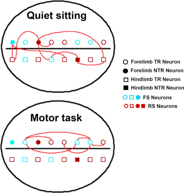
Figure 9.
Significant associations in MI neural networks. Pictorial representation of the pattern of coincident spike activities seen between neurons with different functions during a typical recording session from one hemisphere. The horizontal black line is a simplified representation of the cruciate sulcus, which divides the cat MI anatomically into rostral and caudal 4γ. Each recorded neuron has three attributes indicated by the shape (circle, forelimb representation; square, hindlimb representation), filling (filled, NTR; unfilled, TR), and color (blue, FS; red, RS) of the symbol. In this recording, during quiet sitting we see more varied interactions between NTR and TR neuronal pairs, between FL and HL neurons, and between RS–RS and RS–FS pairs. In contrast, during task performance we see that interactions are limited to those between TR neurons from forelimb representations, and FS–FS or RS–FS pairs.

