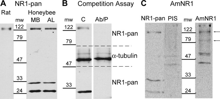
Figure 1.
Detection of the NR1 subunit by Western blot. A, Detection of the NR1 subunit in the MB and the AL regions of the honeybee brain and in rat brain with NR1-pan. B, The specificity of NR1-pan was evaluated in a peptide competition assay, on a honeybee brain extract. The membrane was cut between ∼79 and ∼47 kDa (dashed lines). In the control experiment, the membranes were incubated with NR1-pan or α-tubulin antibodies (C). In the peptide competition assay, the NR1-pan and α-tubulin antibodies were preincubated with the antigenic peptide (Ab/P) before the incubation on the membranes. C, Detection of the NR1 subunit in the honeybee brain with AmNR1. The NR1 subunit was detected with NR1-pan, with the AmNR1 preimmune serum (PIS), and with the AmNR1 serum (AmNR1). Arrows indicate proteins detected with AmNR1 that were not detected with PIS. mw, Molecular weight in kilodaltons.

