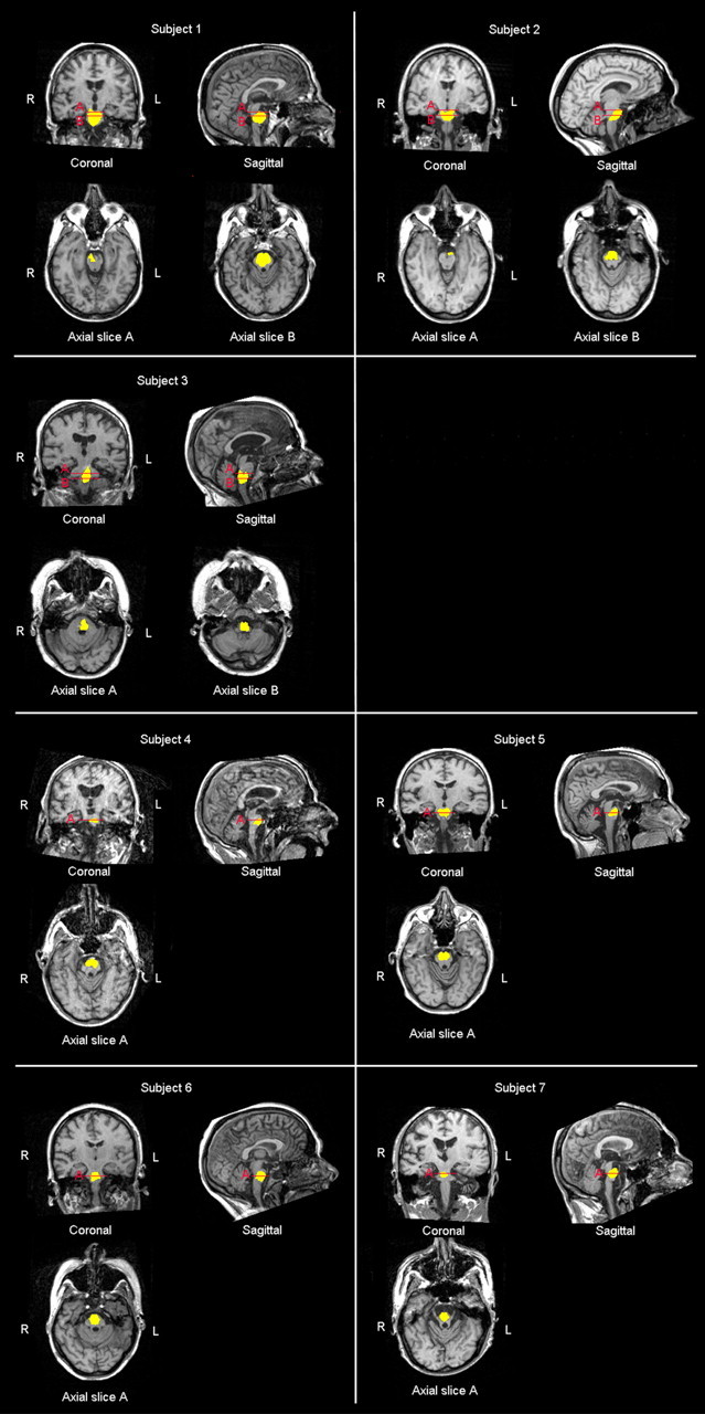
Figure 2.

MR scans showing that subjects 1 and 2 had brainstem lesion including the midbrain and the pons, subject 3 presented a lesion involving the pons and the medulla oblongata, whereas the remaining subjects showed a lesion selectively involving the pons.
