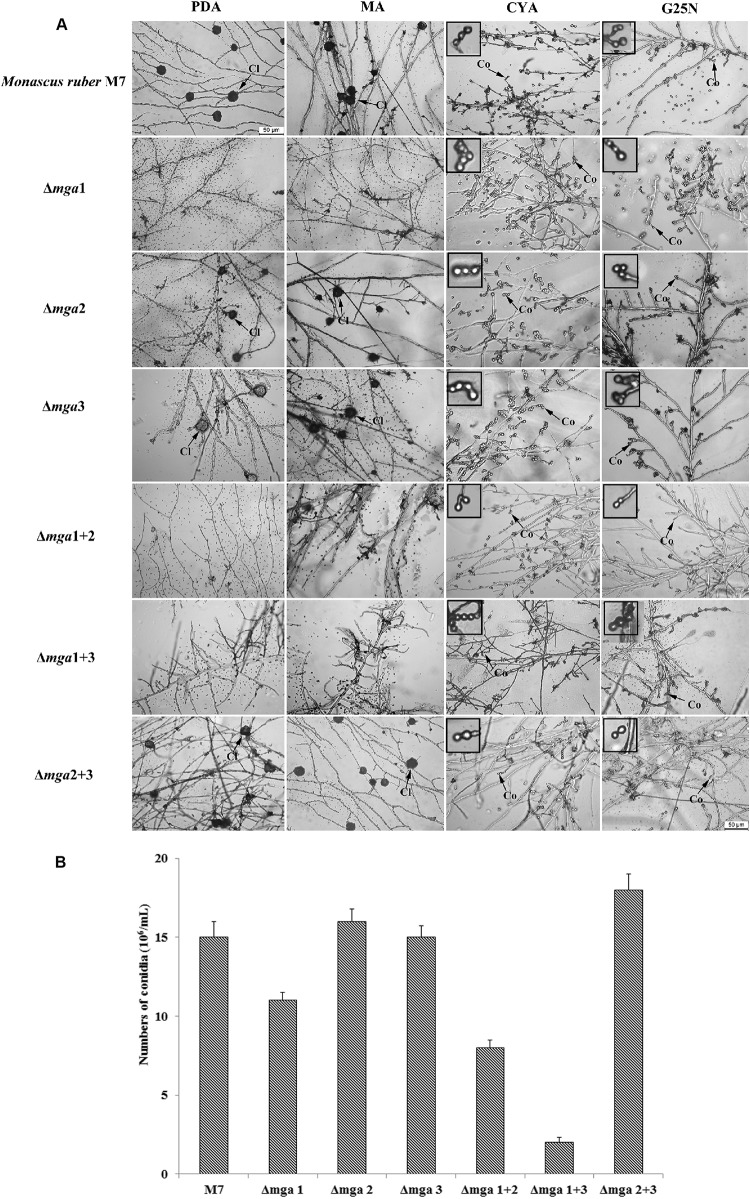
FIGURE 2.
Microscopic structures of Gα mutants and Monascus ruber M7. (A) Cleistothecial (Cl) and conidial (Co) morphologies among M7 and Gα mutants were observed on PDA, MA, CYA, and G25N plates cultured at 28°C for 5 days. The enlarged areas are indicated by arrows. Size bar = 50 μm. (B) The numbers of conidia of the indicated strains were measured after growing on PDA medium at 28°C for 5 days.

