
Figure 2.
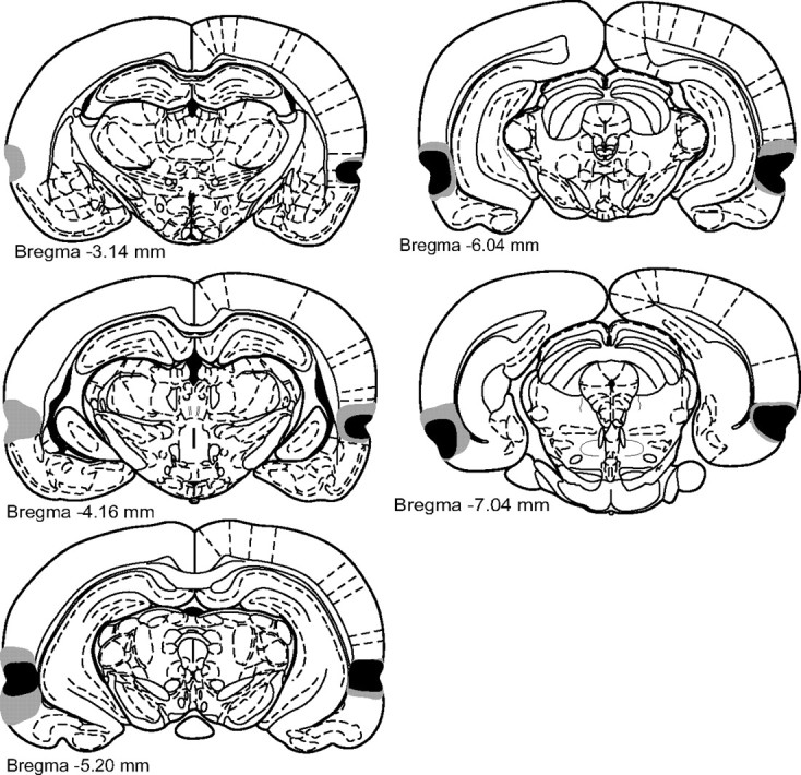
Infusions of the excitotoxin NMDA caused extensive cellular damage throughout PRh. Coronal sections illustrating the extent of the largest (gray) and smallest (black) lesions of PRh, from 3.14 to 7.04 mm posterior to bregma (Paxinos and Watson, 1998), in the bilateral PRh lesion experiment.