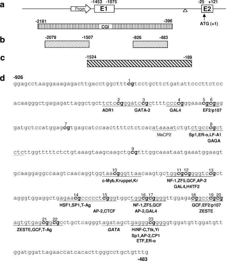
Figure 1.
The SNCA region of interest. a, Schematic drawing of the 5′ region of SNCA with exons 1 and 2 (boxes) and the 5′ UTR and intron 1 (line). The arrow indicates the position of the putative core promoter; the triangle indicates the position of a rudimentary TATA box. Dimension of the CGI is depicted by the stripped box. Numbers are relative to the translation start site (ATG + 1). b, Fragments SNCA(−2079/−1507) (promoter) and SNCA(−926/−483) (intron) were used for bisulfite sequencing. c, Fragment SNCA(−1524/−189) was used for reporter assays. d, Predicted transcription factor binding sites within SNCA(−926/−483) (intron) adjacent to CpG dinucleotides.

