
Figure 1.
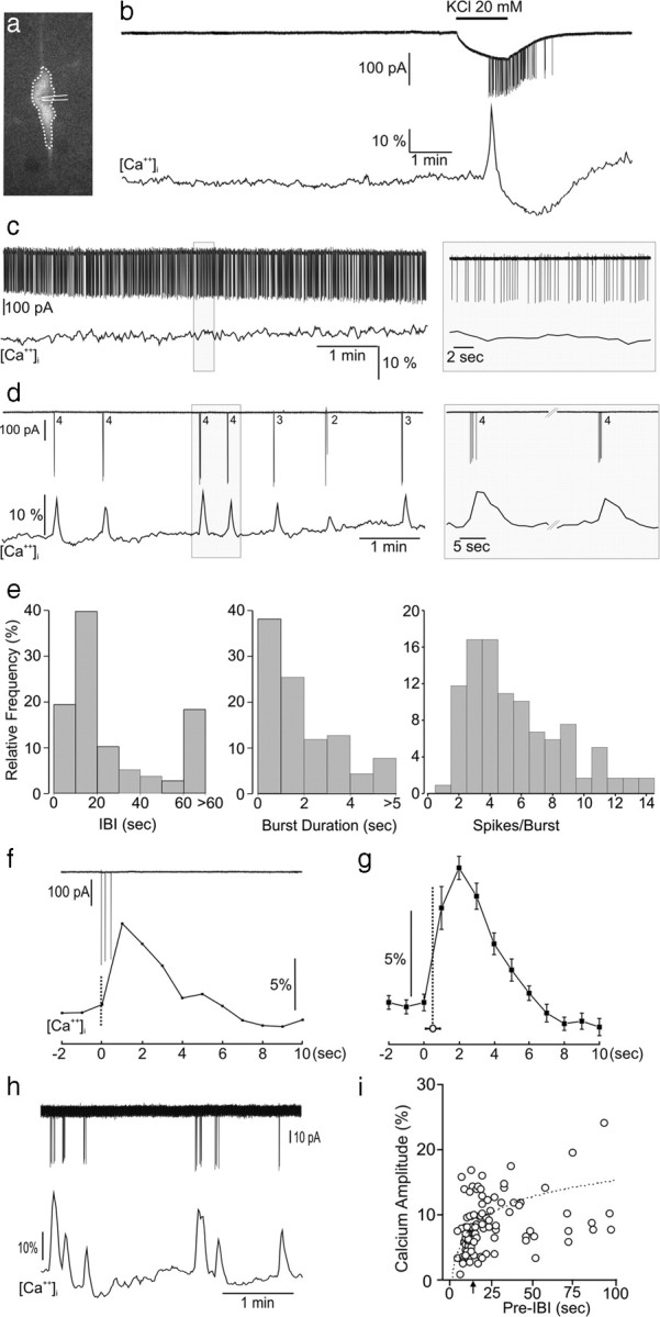
Relationship between action potential spiking and intracellular calcium concentrations [Ca2+]i in adult GnRH neurons. a, Fluorescent GnRH neuron in a brain slice of a GnRH-Pericam transgenic mouse indicating the soma/proximal dendrite area of interest (dotted line) for [Ca2+]i imaging and cell-attached recording electrode. b, Real-time dual electrical (action currents; top)–Ca2+ (bottom) recording from a silent GnRH neuron subsequently challenged with 20 mm KCl that generates a burst of firing and associated Ca2+ transient. c, Dual recording from a continuously active GnRH neuron. d, Dual recording from a burst firing GnRH neuron showing the typical perfect correlation between each burst (numbers = number spikes in burst) and occurrence of Ca2+ transients. e, Parameters of burst firing in GnRH neurons, IBI. f, Higher-temporal-resolution view of relationship between a three-spike burst and Ca2+ transient. g, Average of burst-transient timing by setting Ca2+ datum point immediately before the transient rise (defined as 3 × SD) as t = 0 and determining the time between that and the initiation of the burst (mean ± SEM; vertical dotted line). h, i, A significant correlation existed between IBI and the amplitude of the subsequent Ca2+ transient (r = 0.49, p < 0.001, n = 106; Spearman rank correlation) with transients occurring within ∼15 s (arrow) of the last burst having reduced amplitudes.