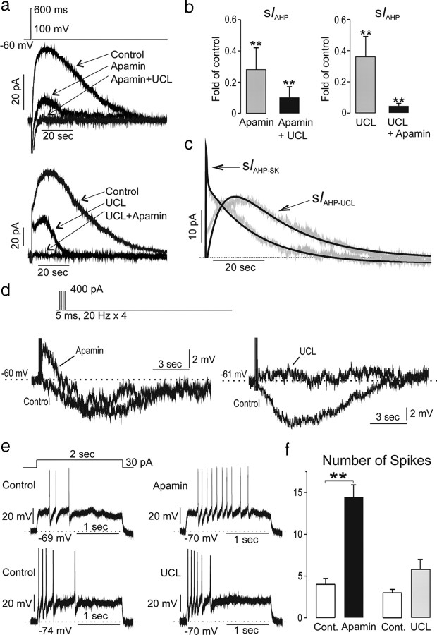
Figure 5.
The AHP in GnRH neurons is mediated by two sIAHPs. a, Perforated-patch, voltage-clamp traces from two GnRH neurons showing the evoked IAHP and its modulation by 300 nm apamin and 10 μm UCL2077. The top cell received apamin then UCL2077 while the bottom cell received the blockers in the reverse order. Each curve is the mean of two repetitions. b, Group (mean ± SEM) data showing fold decrease in IAHP by apamin alone and then combined with UCL2077 (left) or UCL2077 alone and combined with apamin (right; **p < 0.01; Dunnett's multiple-comparison test vs control; n = 5 each group). c, Derived dynamics of apamin- and UCL2077-sensitive currents generated by averaging subtracted curves from individual cells (n = 8). The kinetics of both apamin-sensitive and UCL2077-sensitive currents were well described by exponential rising and decay functions (black lines; with r = 0.89 and r = 0.87, respectively). d, Effects of apamin or UCL2077 on evoked (400 pA for 5 ms at 20 Hz) AHP measured in current-clamp mode. Apamin evoked a large afterdepolarization potential and variable (p > 0.05) suppressive effects on the AHP. UCL blocked the AHP only. e, Effect of apamin (top) and UCL2077 (bottom) on intraburst dynamics induced by 30 pA current for 2 s. f, Group data (mean ± SEM, n = 5 each) showing effects of apamin and UCL2077 on number of spikes per burst. **p < 0.01; Wilcoxon signed-ranks test.

