
Figure 1.
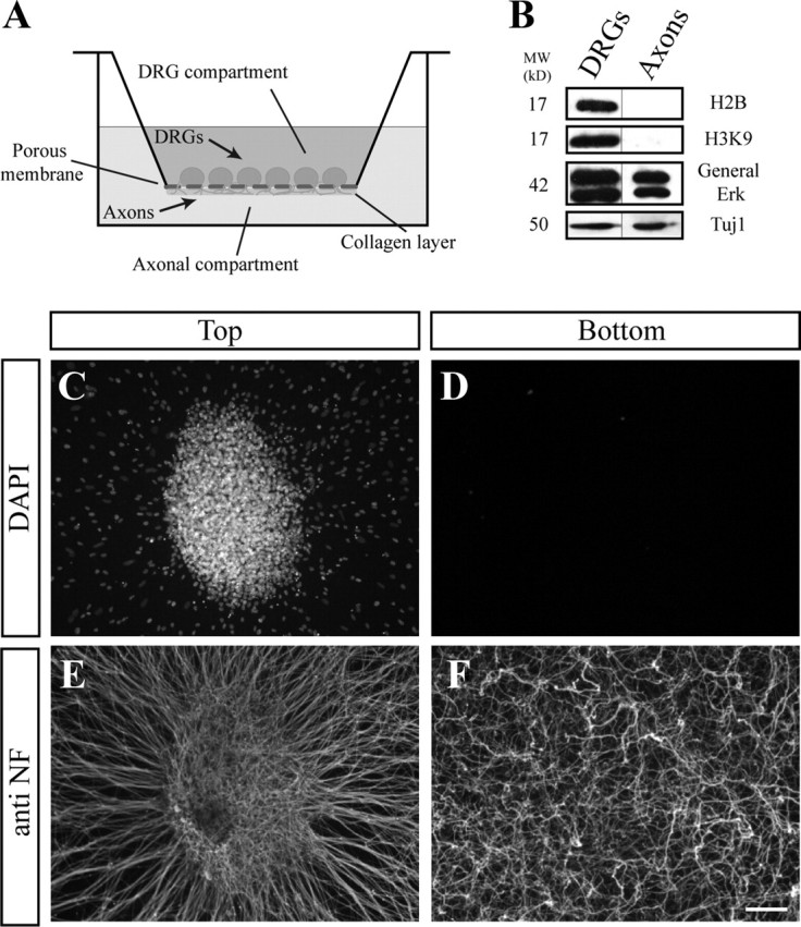
Axonal preparations. A, Schematic illustration of the modified Twiss filter system. C–F, DRG explants were grown on the filter for 48 h, allowing the axons to cross through the 1 μm pores to the bottom compartment and extend within the collagen layer on the bottom side of the membrane. C, E, The upper compartment contains DRG neurons and migrating cells (DAPI) (C) and axons stained with anti-neurofilament (E). In contrast, in the bottom compartment, although numerous axons have crossed the filter (F), no cell bodies are seen (D). Scale bar, 200 μm. B, Biochemical analysis of DRG extracts (left) and axonal preparation (right) using Western blot. The nuclear proteins histones (H2B and H3K9) are present solely in the DRG preparation collected from the upper compartment. Importantly, ERK, which was previously shown to reside in axons, can be detected in our axonal extract.