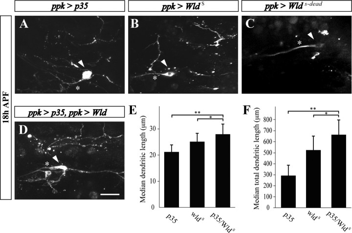
Figure 8.
Cooperation of caspases and the NAD+ pathway in ddaC neuron dendrite pruning. A, B, In flies overexpressing p35 (A) and WldS (B), fragmented ddaC dendritic branches are persisting at 18 h APF. C, However, in the fly strain overexpressing an enzyme dead Wlds (WldS-dead), no ddaC dendritic branches are detected. D–F, Combined expression of both p35 and WldS results in a greater protection than each of them alone. Scale bar, 20 μm. Branch length of ddaC neurons was measured manually within regions of interest using the ImageJ program. Statistical analysis was performed using nonparametrical ANOVA (Kruskal–Wallis). Error bars represent the confidence interval of the median. E, Median dendritic length. F, Median of total dendritic length. *p < 0.05, **p < 0.01.

