
Figure 7.
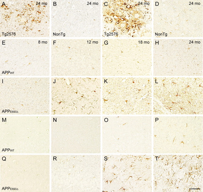
Glial activation in APPE693Δ-Tg mice. A–T, Brain sections of 8 (E, I, M, Q)-, 12 (F, J, N, R)-, 18 (G, K, O, S)-, and 24 (A–D, H, L, P, T)-month-old Tg mice were stained with antibodies to Iba-1 (A, B, E–L) and GFAP (C, D, M–T), which are markers of microglia and astrocytes, respectively. All images were taken from the hippocampal CA3 region, except those of the Tg2576 mice, which were obtained from cerebral cortex. The Tg2576 mice (A, C) at 24 months exhibited massive staining with these antibodies around amyloid plaques, while the non-Tg littermates (B, D) exhibited no staining at 24 months. The APPWT-Tg mice (E–H, M–P) possessed no Iba-1-positive cells and only a few GFAP-positive cells at 24 months. In contrast, the APPE693Δ-Tg mice (I–L, Q–T) displayed Iba-1-positive cells from 12 months and GFAP-positive cells from 18 months in both the hippocampus and cerebral cortex. Scale bar, 30 μm.