Figure 8.
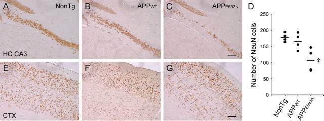
Neuronal loss in APPE693Δ-Tg mice. A–C, E–G, Brain sections of Tg mice were stained with an antibody to NeuN, a marker of mature neurons. Compared with non-Tg littermates (A, E) and APPWT-Tg mice (B, F), APPE693Δ-Tg mice (C, G) exhibited significant decrease in NeuN-positive cells in the hippocampal CA3 region, but no decrease in cerebral cortex at 24 months; hippocampal CA3 region (A–C) and cerebral cortex (E–G). No significant difference was observed between non-Tg littermates and APPWT-Tg mice at 24 months. D, NeuN-positive cells in the pyramidal cell layer of the hippocampal CA3 region were counted within 900 μm from its end toward the dentate gyrus in the photographs. *p = 0.0044 versus NonTg; p = 0.0121 versus APPWT-Tg (n = 4). CTX, Cerebral cortex; HC, hippocampus. Scale bars, 100 μm.

