
Figure 2.
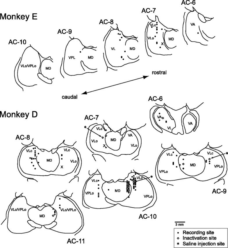
Locations of the task-related neurons and local inactivation. Recording sites were reconstructed from histological sections on the basis of stereotaxic coordinates of electrode penetrations and several electric lesions made during experiments. Black dots indicate the recording sites. White diamonds indicate the sites of muscimol injection. Asterisks indicate the sites of saline injection. The levels of frontal sections are shown as the position posterior to the anterior commissure (AC). VLc and VLo, Caudal and oral part of the ventrolateral nucleus, respectively; VPL, ventroposterolateral nucleus; X, area X.