
Figure 8.
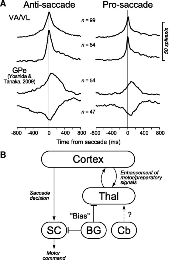
A, Comparison of the population activity for neurons in the motor thalamus and the GPe. The data for anti/prosaccades are shown separately in different columns. On each column, the top trace shows the population activity computed for all task-related neurons recorded from the motor thalamus. The second trace was computed only for the neurons in the motor thalamus that lacked a significant firing modulation during the instruction period. The bottom two traces plot the data of GPe neurons that exhibited either increased (third trace) or decreased (fourth trace) firing rate during saccades obtained in the previous study (Yoshida and Tanaka, 2009). B, A possible neural mechanism of the basal ganglia-thalamocortical pathways in the generation of antisaccades. In this diagram, the motor drive and preparatory signals for antisaccades are processed within the mutual thalamocortical projections, while the basal ganglia regulate the gain of this intrinsic pathway. The arrows and bars indicate excitatory and inhibitory connections, respectively. BG, Basal ganglia; Cb, cerebellum; Thal, thalamus.