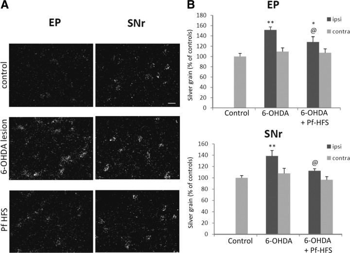
Figure 10.
A, B, Photomicrographs (A) and quantitative analysis (B) showing the effects of separate or combined unilateral 6-hydroxydopamine lesion and Pf-HFS on COI mRNA expression in EP and SNr. Sections were processed for in situ hybridization with 35S-radiolabeled COI oligonucleotide probe and emulsion autoradiography. The data presented in the graphs are the means ± SEM of silver grain number determined from n animals per condition and are expressed as percentages of controls. Statistical comparison was performed using a one-way ANOVA followed by Student–Newman–Keuls test. Scale bar, 20 μm. **p < 0.01 and *p < 0.05 compared with control values; @p < 0.05 compared with 6-OHDA lesion values.

