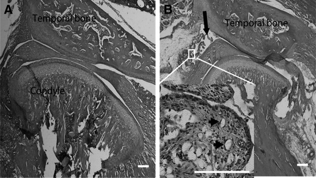
Figure 2.
Pathological examination of CFA-induced TMJ inflammation. A, Photomicrograph of TMJ injected with saline for 24 h. B, Photomicrograph of TMJ injected with CFA for 24 h. Exuberant hypertrophy of the synovial tissue (enlarged panel), angiogenesis (indicated by arrowheads), and fibrin-like exudates (indicated by arrow) in the joint space were observed. Scale bar, 200 μm.

