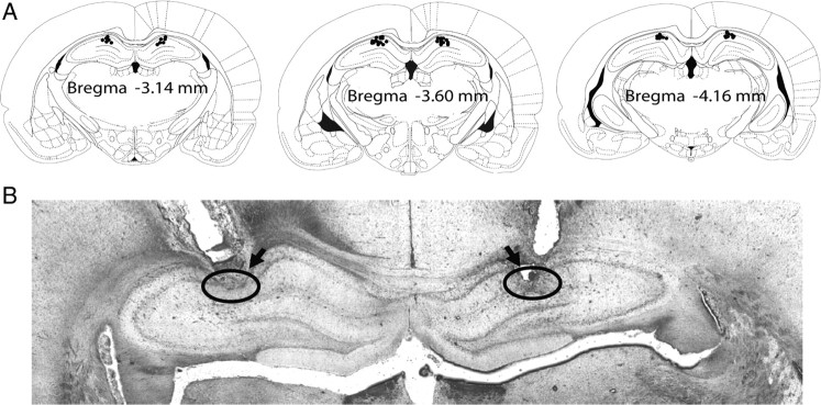
Figure 6.
Confirmation of cannula placements in the hippocampus in rats. A, Reconstruction of serial coronal sections of the brain illustrates the bilateral injection sites of the cannulas. Black dots on the schematic illustration of the coronal section of rat brain [adapted from the rat brain atlas of Paxinos and Watson (1997)] indicate the placements of cannulas in the brain. B, Representative microphotograph of the coronal section shows the placements (indicated by circles and arrows) of a pair of cannulas in the CA1 region of the hippocampus.

