
Figure 5.
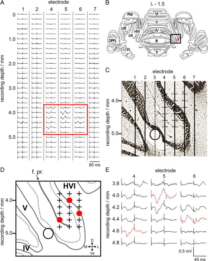
Example single penetration of a microelectrode array in an anesthetized rabbit. A, Evoked LFP activity from ipsilateral periocular stimulation shown for every recording site, for each of the six recording microelectrodes in the array (#1, #2, #4–7; averages of 20 trials). At each step, the array was advanced 200 μm. Two low-intensity electrical stimuli (see Materials and Methods, Electrical stimulation) were delivered 40 ms apart, the timing of which can be seen from the positions of the stimulus artifacts. A box highlights the region with periocular-evoked climbing fiber activity consistent with a periocular microzone; these traces are expanded in E. These tracks are 4.3, 4.6, and 4.9 mm lateral to the estimated midline. Bilateral forelimb and vibrissal stimulation did not evoke climbing fiber activity in this region (data not shown). C shows an expanded histological reconstruction of the estimated path of the electrodes (vertical lines) based on the presence of electrolytic lesions (open circle) made using the lesion-making electrode (#3); the location of this portion of the reconstruction represented on an annotated standard coronal section (at 1.5 mm caudal to lambda; λ − 1.5) is illustrated by the box in B. Red circles in B indicate the estimated location of the largest-amplitude periocular CFP in each electrode track. An annotated diagram of the region shown in C, labeling the lobules, primary fissure, and anatomical directions is drawn in D. The estimated recording locations of the evoked LFPs shown in the box in A and expanded in E are highlighted with plus signs. Red circles in D represent the locations of the largest-amplitude periocular-evoked CFPs on each of the electrode tracks (red traces in E). These points are also projected on to the standard coronal section in B. PM, Paramedian lobule; cr I, crus I; cr II, crus II; DPFL, dorsal paraflocculus; FL, flocculus; f. pr., primary fissure; D, dorsal; Ve, ventral; M, medial; L, lateral.